Smart Medical Image Analysis for Chest CT for research purpose only
2020-09-17
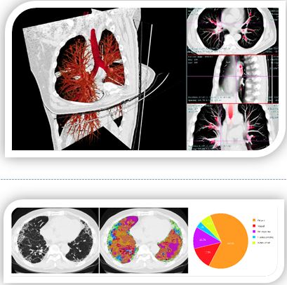
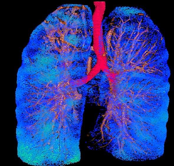
A total solution for comprehensive analysis of Chest CT image: revealing the lung nodule characterization as well as the diffuse lung disease. Facilitating radiologists in disease diagnosis, treatment planning, therapy monitoring and follow-up. The qualitative and quantitative results can be used for academic publications and clinical cross reference; Through advanced visualization solution, the follow-up of nodules is no longer a subjective measurement on particular image, but an objective measurement of volumetric, structural and textual variations. Advanced analytic platform improves precision in clinical practice.
CT Image Viewer
Automated anatomical labelling
2D, 2.5D, 3D, 4D image reconstruction
File export for quantification data
Batch analysis for multiple dataset
Multiple annotating functions
Multiple measuring functions
Model file export for 3D printer
Customized Image Analysis Module
Clinical collaboration
Image correlational study
Longitudinal image analysis
Algorithm development and licensing
Anatomical Image Analysis
Coronary artery: analysis for stenosis and plaque compositions
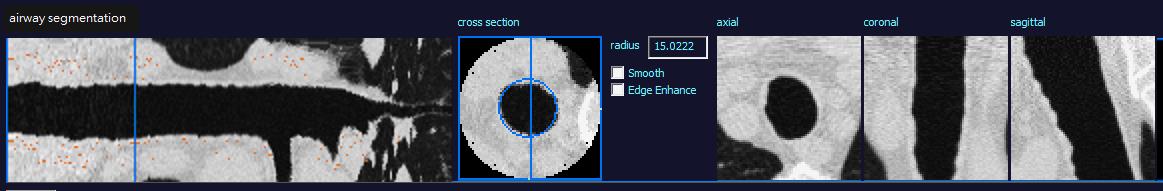
Pulmonary airway: wall thickness, lumen diameter ratio
Emphysema: severity quantification and lung function correlational analysis; parametric response map
Lung nodule detection: morphology and texture analysis